X-Ray Home
Helpful Links
About the Facility
The NCMN X-Ray Structural Characterization Facility is dedicated to materials identification and characterization through non-destructive, X-Ray Diffraction (XRD) technique. The specific applications include Powder diffraction, x-ray reflectometry, small angle scattering, pole figure, reciprocal space mapping, Grazing incidence in-plane diffraction, x-ray crystallography etc. Non-ambient powder and single crystal diffraction is also available at selected temperature range.
Powder diffraction is routinely used for phase identification, finding relative ratio of phases, structural phase transitions, percentage of crystallinity analysis of partially crystalline materials, etc. Advanced hardware and high resolution optics are available to investigate thin films, multilayers, highly oriented and epitaxial thin films. Grazing incidence in-plane diffraction provides valuable information of the structural order in the plane of the film. X-ray crystallography is unique in capabilities to unravel the absolute crystal structure from single crystal diffraction.
The following instruments are available at the NCMN X-ray Scattering Facility: (1) Rigaku SmartLab Diffractometer (2) Bruker D8 Discover Diffractometer (3) PANalytical Empyrean Diffractometer (4) Rigaku D-Max/B Diffractometer (5) Rigaku Multiflex diffractometer and (6) Bruker Photon 100 Single Crystal diffractometer.
Acknowledgement Text
Agencies including NSF and the University providing partial support of our Nebraska Nanoscale Facilities and NCMN Facilities require that the following words be included at the end of any Acknowledgement section of a paper in which experimental work was done in NNF-NCMN facilities:
The research was performed in part in the Nebraska Nanoscale Facility: National Nanotechnology Coordinated Infrastructure and the Nebraska Center for Materials and Nanoscience (and/or NERCF), which are supported by the National Science Foundation under Award ECCS: 2025298, and the Nebraska Research Initiative.
Diffractometers

1. Rigaku SmartLab Diffractometer
This is a state-of-the-art instrument including a dedicated arm for detector movement in-plane and perpendicular to the sample surface, thereby able to probe structure in both directions without tilting the sample. The instrument is equipped with cross beam optics device hardware that will allow switching between focussing (BB) and the parallel beam (PB) geometries easily. SmartLab guidance software helps the optics and sample alignment automated and prepares optimum scan conditions. SmartLab can be configured for grazing-incidence in-plane XRD, grazing-incidence XRD, x-ray reflectivity, high-resolution XRD (rocking curves, reciprocal space maps), texture (pole figures), residual stress analysis and small angle x-ray scattering study of nanoparticles. The diffractometer uses Cu Kalpha radiation (wavelength of about 1.54 Å).
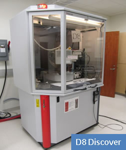
2. Bruker-AXS D8 Discover Diffractometer
This also is state-of-the-art machine including Vantec-500 area detector, centric 1/4-circle Eulerian cradle, domed hot stage, hi-flux in-plane hardware, laser/video sample-alignment system, Göbel mirror, fine tilt stage, and dual-beam path analyzer module. The system can be configured for grazing-incidence in-plane XRD, grazing-incidence XRD, x-ray reflectivity, high-temperature XRD, high-resolution XRD (rocking curves, reciprocal space maps), texture (pole figures), residual stress, and capillary diffraction. Bruker D8 Discover diffarctometer is configured in parallel beam geometry with Cu Kalpha radiation (wavelength of about 1.54 Å).
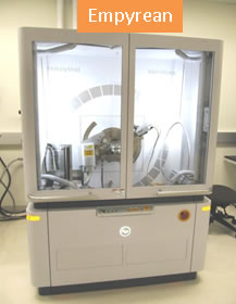
3. PANalytical Empyrean Diffractometer:
Empyream is a dedicated powder diffractometer with advanced detection capabilities. It is equipped with PIXcel 3D detector that can be operated with 0D, 1D and 2D detection capabilities. This instrument consists of 3 kW copper target and theta-theta goniometer. The samples can be placed in zero-background holder and the holder remains horizontal as well as it can be spun continuously during the scan.
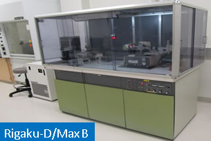
4. Rigaku D/Max-B Diffractometer:
X-Rays are produced by a 1.8 kW, sealed tube Cobalt target. The diffracted beam then converges (is “focused”) into a diffracted beam monochromator which removes all radiation except the Co Kalpha wavelength (about 1.7903 Å) which then enters a scintillation counter. The sample and detector are rotated with respect to the incident beam at angles theta and 2theta respectively. A typical XRD scan consists of a plot of detector angle (2theta) vs. diffracted intensity. These diffractograms can be printed out or saved to disk in application-friendly ASCII format.
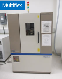
5. Rigaku Multiflex Diffractometer:
This instrument consists of 2 kW copper target and theta-theta goniometer. The sample holder remains horizontal during the scan and therefore, there is no need to use adhesive substances to mount samples on to a low background sample holder plate. Similar to D/max-B, Rigaku Multiflex is also configured in focusing geometry where a secondary monochromator removes the scattered signal except that corresponding to Cu Kalpha wavelength. Pre-aligned sample holder and friendly operating software makes the powder diffraction experiments very easy with this instrument.
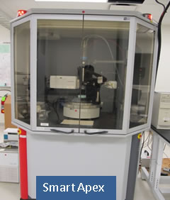
6. Bruker Photon 100 Single Crystal Diffractometer:
Smart Apex Single Crystal Diffractometer is a dedicated instrument for Crystallography studies (absolute structure determination using a single crystal sample). This instrument delivers intense, monochromatic beam of Mo Kalpha radiation (0.7107 Å) produced using Graphite monochromator from a sealed Mo X-ray tube and collimated with a pinhole collimator. The single crystal is mounted on the tip of a glass fiber or a nylon loop affixed to a Goniometer head will be attached to a fixed χ, 3-axis goniometer. The sample is optically aligned using Video camera and a manual control module. The sample temperature can be varied from 100 – 500 K using Cryostream controller (Oxford Instruments). The detector (Photon 100 CMOS detector) is placed at about 5 – 6 cm from the sample. The frame buffer computer is provided with APEX III software for sample centering, data collection (sample unit cell determination, and detailed structure determination), data integration, absorption correction, and structure determination and refinement.
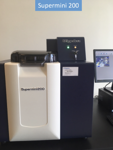
7. Rigaku Supermini200 WDXRF Spectrometer:
Supermini 200 is a benchtop sequential wavelength dispersive X-ray fluorescence (WDXRF) spectrometer with high resolution and lower limits of detection for elemental analysis of Fluorine (F) through Uranium (U). The 200 W, air cooled, Pd target operated at 50 kV and 4 mA produces the excitation spectrum capable of detecting light elements faster. It uses a 3-crystal changer analyzing unit that supports the standard LiF (200) as well as PET and RX25 crystals that works efficiently in the within the elemental range of U - F. Two detectors are being used – a gas flow proportional counter (F-PC) suited for light elements and Scintillation counter (SC) suited for heavy elements. F-PC uses P-10 (UHP grade 90 % Argon and 10 % Methane) gas flow for operation. Samples are placed in a 12-position sample turret and are automatically loaded to the optical chamber with software control. The maximum dimensions of the sample are 44 mm in diameter, and 33 mm in height. The samples can be in the form of powders, bulk solids or liquids. Spectrometer chamber environment is usually vacuum when analyzing bulk samples, however, the environment can be set to helium for liquid samples. The sample also can be spun during the measurement. Characteristic emission lines detects the elements present and the intensity analysis using Fundamental Parameter method determines the chemical composition.
Data Reduction and Analysis
Various XRD data reduction and analysis software from Rigaku, Bruker and PANalytical as well as molecular graphical visualization and analysis tools are available for the users of the facility. NCMN subscribes structural databases from International Center for Diffraction Data (ICDD) PDF–4+ and Cambridge Structural Database (CSD) for the up to date information of inorganic and organic materials.
Facility Contacts

Ather Mahmood
Facility Specialist - Primary
006A Jorgensen Hall(402) 472-3693
ather.mahmood@unl.edu

Bibek Tiwari
Facility Specialist - Secondary
006A Jorgensen Hall(402) 472-3693
tiwaribibek@huskers.unl.edu